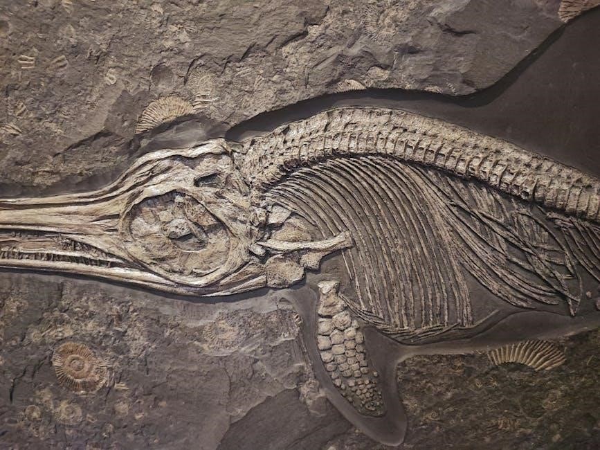
pdf of skeletal system
Overview of the Skeletal System
The skeletal system consists of 206 bones, cartilage, ligaments, and tendons, providing structural support and protection for the body․ It is divided into axial and appendicular sections․
1․1 Definition and Importance
The skeletal system is a framework of 206 bones, cartilage, ligaments, and tendons that provides structural support, protects vital organs, and facilitates movement․ It plays a crucial role in storing minerals like calcium and phosphorus and is the site of blood cell production in the bone marrow․ This system is essential for maintaining posture, enabling mobility, and safeguarding internal organs such as the brain, heart, and lungs․ Its functions are vital for overall health and bodily functions․
1․2 Components of the Skeletal System
The skeletal system comprises 206 bones, cartilage, ligaments, and tendons․ Bones provide structural framework, while cartilage cushions joints, reducing friction․ Ligaments connect bones, stabilizing joints, and tendons attach muscles to bones, enabling movement․ The system also includes bone membranes and marrow, which produce blood cells․ These components work together to form a dynamic structure essential for body support, protection, and mobility, ensuring proper bodily functions and overall health․
Functions of the Skeletal System
The skeletal system provides structural support, protects internal organs, facilitates movement through muscle attachments, and stores essential minerals like calcium․ It also produces blood cells․
2․1 Support and Framework for the Body
The skeletal system acts as the body’s structural framework, providing support and maintaining posture; Bones of varying sizes and shapes form a network connected by joints, enabling flexibility and movement․ Ligaments and tendons stabilize these bones, allowing the body to function efficiently․ This framework is crucial for daily activities, ensuring stability and enabling a wide range of motions essential for life․
2․2 Protection of Internal Organs
The skeletal system plays a vital role in protecting vital organs․ Bones such as the skull safeguard the brain, while the rib cage shields the heart and lungs․ Vertebrae protect the spinal cord, ensuring the nervous system remains intact․ This protective function is essential for maintaining overall health and preventing damage to critical body systems․
2․3 Facilitating Movement
The skeletal system enables movement by acting as a framework for muscle attachment․ Bones, joints, and ligaments work together to allow mobility․ Joints, classified as synarthroses, amphiarthroses, and diarthroses, permit varying degrees of movement․ This system supports both voluntary actions, like walking, and involuntary movements, ensuring dynamic interaction between bones and muscles for optimal mobility and flexibility in daily activities․
2․4 Storage of Minerals and Blood Cell Formation
Bones serve as reservoirs for essential minerals like calcium and phosphorus, which are vital for various bodily functions․ Additionally, bone marrow within the skeletal system produces blood cells, including red blood cells, white blood cells, and platelets, crucial for oxygen transport, immune defense, and blood clotting․ This dual function highlights the skeletal system’s role beyond structure, contributing to metabolic balance and hematopoiesis, ensuring overall health and bodily function․
Axial and Appendicular Skeleton
The skeletal system is divided into axial and appendicular parts․ Axial includes skull, spine, ribs, and sternum, while appendicular comprises limbs and their connecting girdles․
3․1 Axial Skeleton
The axial skeleton includes the skull, vertebral column, ribs, and sternum․ It forms the body’s central framework, providing protection for vital organs like the brain and heart․ The skull consists of cranial and facial bones, while the vertebral column supports the spinal cord․ Ribs attach to the spine and sternum, creating a protective cage for the chest cavity․ This system ensures structural stability and safeguards internal organs from injury or damage․
3․2 Appendicular Skeleton
The appendicular skeleton comprises the upper and lower limbs, shoulders, hips, and pelvic girdles․ It connects to the axial skeleton, facilitating movement and supporting the body’s appendages․ The upper limb includes the arm, forearm, and hand bones, while the lower limb includes the thigh, leg, and foot bones․ This system enables locomotion, manipulation of objects, and transfer of forces from the body to the environment, essential for daily activities and mobility․

Types of Bones
Bones are classified into four types: long, short, flat, and irregular․ Each type has distinct functions, such as support, protection, and movement, based on their shapes․
4․1 Long Bones
Long bones are elongated and cylindrical, with growth primarily occurring in length․ They have a shaft (diaphysis) and two ends (epiphyses), which are wider for joint articulation․ Examples include femur, humerus, tibia, and fibula․ These bones provide structural support and facilitate movement by serving as attachment points for muscles, enabling activities like walking and running․ Their structure allows for strength and flexibility, making them crucial for locomotion and overall mobility in the human body․
4․2 Short Bones
Short bones are cube-like in shape, providing stability and limited movement․ Found in the wrists (carpals) and ankles (tarsals), they absorb shock and distribute weight․ Their flat surfaces allow for articulation with adjacent bones, enabling limited but essential movements․ Short bones are dense and sturdy, offering support while maintaining flexibility in joints․ They play a vital role in the skeletal system by facilitating balance and stability in the body’s structure․
4․3 Flat Bones
Flat bones are thin and plate-like, serving primarily for protection and providing surfaces for muscle attachment․ Examples include the skull bones, sternum, and ribs․ These bones are strong yet lightweight, offering defense for internal organs like the brain and heart․ Their broad surfaces allow for extensive muscle attachment, aiding in movement and stability․ Flat bones are essential for safeguarding vital structures while contributing to the body’s overall framework․
4․4 Irregular Bones
Irregular bones have unique, complex shapes that enable specialized functions․ Examples include vertebrae, which support the spinal column, and facial bones, which form the structure of the face․ These bones often have multiple surfaces for muscle and ligament attachment, enhancing movement and providing protection for vital organs․ Their intricate designs allow for a wide range of functions, from supporting the body’s weight to enabling facial expressions and safeguarding internal structures․

Joints and Their Classification
Joints are points where bones connect, allowing movement and stability․ They are classified into synarthroses (immovable), amphiarthroses (slightly movable), and diarthroses (freely movable) based on motion capability․
5․1 Synarthroses (Immovable Joints)
Synarthroses are immovable joints that provide stability and support․ Examples include sutures in the skull and the joint between the hyoid bone and thyroid cartilage․ These joints are essential for protecting vital structures, such as the brain, by preventing movement in critical areas․ They are typically fibrous, with dense connective tissue holding bones together, ensuring rigidity and structural integrity without compromising the body’s ability to function effectively in other movable regions․
5․2 Amphiarthroses (Slightly Movable Joints)
Amphiarthroses are joints that allow limited movement, providing flexibility while maintaining stability․ Examples include intervertebral discs and the pubic symphysis․ These joints are connected by fibrocartilage, enabling slight movements such as gliding or bending․ They are crucial for absorbing shock and distributing stress in areas like the spine, while still offering structural support․ This type of joint balances mobility and rigidity, ensuring proper function in regions requiring moderate movement without compromising overall stability․
5․3 Diarthroses (Freely Movable Joints)
Diarthroses are freely movable joints that enable a wide range of motion․ Examples include shoulder, hip, and knee joints․ These joints are characterized by a synovial cavity filled with synovial fluid, reducing friction between bones․ Articular cartilage covers the ends of bones, allowing smooth movement․ Ligaments and muscles provide stability while maintaining flexibility․ Diarthroses are essential for activities like walking, running, and grasping, enabling complex movements that are vital for daily functions and maintaining an active lifestyle․

Common Skeletal Injuries and Fractures
The skeletal system is prone to injuries like fractures, which can be transverse or oblique, affecting the bone structure and its protective functions․
6․1 Types of Fractures
Fractures are classified based on their severity and pattern․ They include transverse fractures, which cut straight across the bone, and oblique fractures, characterized by diagonal breaks․ Additionally, stress fractures occur due to repetitive strain, while compression fractures often affect the vertebrae․ Each type requires specific treatment to restore bone integrity and function․ Understanding these classifications aids in proper diagnosis and care for skeletal injuries․
6․2 Transverse and Oblique Fractures
Transverse fractures occur straight across the bone, typically resulting from a direct blow or stress․ Oblique fractures are diagonal, often caused by bending or twisting forces․ Both types disrupt bone continuity and require immobilization for healing․ Treatment may involve casting, surgery, or physical therapy to restore strength and mobility․ Proper diagnosis and care are essential to prevent complications and ensure full recovery from these common injury types․

Skeletal System Resources
Explore comprehensive PDF guides, 3D models, and interactive tools for detailed skeletal system study․ Resources include educational documents, visual aids, and apps for enhanced learning experiences․
7․1 PDF Guides and Documents
Downloadable PDF guides provide in-depth insights into the skeletal system, covering bone structure, functions, and detailed anatomical illustrations․ These documents are ideal for students and researchers seeking comprehensive knowledge․ They often include sections on axial and appendicular skeletons, joint classifications, and common injuries; Available for free or purchase, these resources are accessible through various educational platforms and websites, offering a convenient way to study the skeletal system in detail․
7․2 3D Models and Visualizations
3D models and visualizations offer interactive and detailed representations of the skeletal system, enabling users to explore bone structures from various angles․ These models are available in formats like STL, OBJ, and FBX, suitable for educational and professional use․ They provide insights into complex anatomical features and are accessible through apps and websites, making them invaluable tools for learning and reference in anatomy and healthcare fields․
